What is laminitis?
 Laminitis, also known as 'founder' is considered the second biggest killer of horses after colic. Whilst not necessarily fatal, horses with severe, acute laminitis are often euthanised due to the amount of pain they are in.
What is Laminitis?
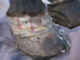
Laminitis is a condition of the foot, but the cause is actually elsewhere in the body. Laminitis affects the laminae within the foot. The easiest way to imagine this is as a Velcro layer between the hoof wall and the coffin bone that holds these two structures together. Laminitis is inflammation and loss of structure of this 'Velcro' layer. As this bond weakens the force of weight and movement begin to rotate the coffin bone (the lowest bone in the foot) out of alignment: in extreme cases the coffin bone can penetrate right through the sole of the foot. As the coffin bone rotates, blood vessels get squeezed and cells within the foot get starved of oxygen and nutrients and start to die. Once this stage has been reached, abscessing is common, as the body tries to expel necrotic material.
Not all cases of laminitis are this extreme. Sometimes horses have only mild laminitis and show no clinical lameness, only subtle deformities in their hooves and a loss of athletic performance. Laminitis can be acute (sudden onset where it is clear that there is something very wrong with your horse) or chronic, an ongoing issue which may have been unrecognised for a long period of time, or that developed insidiously from low grade or even sub-clinical laminitis, or that has evolved from an acute case that was not dealt with immediately or very successfully.
Horses with metabolic disorders such as insulin resistance are often susceptible to chronic laminitis. Whilst on the surface a horse with mild chronic laminitis may appear to be managing, the adverse pressure on the sensitive structures and tissues of the foot are still there. Over time even the coffin bone itself can start to be eroded away because of this adverse pressure. The longer these structures are subject to damage the less likely it is for the horse to recover full soundness.
WHAT CAUSES LAMINITIS?
Laminitis manifests itself in the foot of the horse, but this is a symptom - the cause of laminitis is elsewhere in the body. The most common cause is too much sugar: the horse that gets into the feed shed and gorges on oats; or the pony that spends too long out on rich pasture. This excess of sugar in the hind gut affects the balance of bacteria: bacteria die releasing toxins, the wall of the bowel becomes leaky and these toxins are released into the bloodstream, triggering laminitis.
Laminitis can be triggered by other illnesses such as retained placenta or pneumonia, and can also be triggered by snake bite.
SYMPTOMS
Symptoms can include high heart rate, a bounding pulse in the lower leg, rapid breathing, fever, sweating, colic, diarrhoea, or depression. The horse is likely to exhibit foot pain and lameness, especially in acute cases, which may range from shifting weight from one foot to another, a 'laminitic stance' in which a horse shifts his weight into his hind feet to relieve the pain in his front feet, to a refusal to stand at all because of the intence pain.
IF YOU ARE CONCERNED THAT YOUR HORSE MAY BE SUFFERING FROM LAMINITIS CONSULT YOUR VETERINARIAN IMMEDIATELY.
DON'T FORGET GOOD NUTRITION
Animals suffering from laminitis are usually put on a carefully controlled diet to restrict the amount of sugar, but the horse still needs essential nutrients, particularly when the body is trying to heal itself. HOOF SUPPORT can be given to animals with laminitis, and contains the proteins, vitamins and minerals the body needs to repair the damage from the inside as new hoof grows down.s
Another very useful supplement is DEVIL's CLAW, nature's anti-inflammatory to help manage the pain and inflammation, particularly over the longer term.
|
FOR THOSE WHO WANT THE SCIENCE:
A common cause of laminitis is carbohydrate overload. Structural carbohydrate, such as pasture, is broken down in the hindgut of the horse through microbial fermentation. A sudden excess of sugar in the diet, such as grain overload or too much rich spring grass, breaks down the balance of this microbial fermentation, triggering laminitis. The excess sugar undergoes rapid microbial fermentation to lactic acid in the normally neutral conditions of the equine hind gut. As more sugar rich feed arrives in the hind gut, conditions become more acidic, with hindgut pH dropping to as low as 4. These acidic conditions favour streptococci bacteria, and the result of the acidic conditions is a population explosion of this particular species. The low pH in the hindgut results in the death of large numbers of other species of bacteria, whose cell walls breakdown releasing endotoxins, exotoxins and microbial DNA into the hindgut. Within the hindgut, further degenerative changes take place. The mucosal barrier of tightly knit epithelial cells that line the glands and lumen of the bowel wall is damaged, with widespread desquamation and sloughing of these epithelial cells. The result is a leaky bowel through which lactic acid, toxins and laminitis trigger factors can pass into the bloodstream. The exact laminits trigger factor has yet to be identified, but Professor Chris Pollitt theorises that the trigger factor disrupts the process of remodelling enzymatic matrix metalloproteinases (MMP) in the hoof: excess MMP is released and activated, resulting in the destruction of laminar attachment. During acute laminitis, a distinct degeneration at the cellular level can be mapped. Initially the secondary epidermal lamella (SEL) change shape: the tips of the SEL change from being rounded to being longer, thinner and pointed. At the same time, the position and shape of the basal cells within the SELs change: from oval to rounded, and from a position away from the basement membrane to abnormally close. At this stage, the secondary dermal lamella (SDL) still interdigitate between the SELs, and their tips are close to the primary epidermal lamella as in a healthy hoof. However, the basement membrane has lifted away from the underlying basal cells at the tapered tips of the SELs. From this point, each time the foot is weighted, the basement membrane slips further away, as it is no longer attached to the basal cells. It also retracts from between the SELs, taking connective tissue with it. In acute laminitis, where the horse is severely lame, the degradation and disorganisation continues at a cellular level. The SELs become a disorganised, amorphous mass, with isolated remnants of basement membrane, but with no effective attachment to any connective tissue. The lamellar tips slide away from their basement membrane connective tissue attachments. This occurs first microscopically, but eventually it can be measured by the naked eye on a radiograph. This is the 'sinker' phase of acute laminitis as attachment fails, and the distal phalanx rotates downwards through weight and movement. |
|
RELATED PRODUCTS |
|||
 |
 |
 |
 |
| Laminitis Recovery | Hoof Builder |
Devil's Claw |
Hoof Support |